 Reviewed by Mr. Ranjeet Kumar
Sr. Audiologist, Speech Therapist & Cochlear Implant Specialist, BASLP on
Reviewed by Mr. Ranjeet Kumar
Sr. Audiologist, Speech Therapist & Cochlear Implant Specialist, BASLP on

Cholesteatoma is a unique disease of the ear in which a skin cyst or sac grows into the middle ear and mastoid. The cyst is not cancerous but can erode tissue and cause the destruction of the ear.
You can purchase the latest hearing aids at a fair price through HearingSol, If you have a query regarding Cholesteatoma and Hearing Loss, feel free to call us at +91-9327901950. We are always here to help you.
What Is Cholesteatoma?
A cholesteatoma is an abnormal, noncancerous skin growth that can develop in the middle section of your ear, behind the eardrum. It may be a birth defect, but it’s most commonly caused by repeated middle ear infections.
It is an abnormal skin growth where a sack of dead skin cells is formed in the middle ear or skin cyst trapped behind the eardrum or the bone behind the ear.
Over time, the skin collects and causes problems like infection, drainage, and loss of hearing.
The skin may take a long time to accumulate and can spread to the area behind the eardrum, or to the bone behind the ear (called the mastoid bone). It can cause an unpleasant smelling discharge followed by the loss of hearing.
How Does Cholesteatoma Occur?
- Causes of Cholesteatoma
There are different reasons as to why cholesteatoma may develop in a person. Repeated infections are one of the most important causes of Cholesteatoma.
Besides ear infections, there may be several reasons behind the cholesteatoma such as: Congenital and Acquired.
Thus the cholesteatoma can be categorized into two types :
- Congenital Cholesteatoma
- Acquired Cholesteatoma
1. Congenital Cholesteatoma
Cholesteatoma can be a birth defect, also called congenital cholesteatoma in medical terminology, and is more commonly occurred as a result of chronic ear infection.
Congenital cholesteatomas can form in the middle ear or in other areas of the ear. This type of cholesteatoma is very rare and occurs when squamous epithelium residue remains in the middle ear during the development of the embryo. The tympanic membrane is intact.
This type of Cholesteatoma fulfill the following conditions :
- Mass media to the tympanic membrane
- Normal tympanic membrane fulfill
- No previous history of ear discharge, perforation or ear surgery
2. Acquired Cholesteatoma
- Symptoms of Cholesteatoma
It is the most common type of cholesteatoma caused by pathological alteration of the eardrum leading to the accumulation of keratin within the middle ear. The causes of acquired cholesteatoma are :
- Eustachian Tube Dysfunction
- Ruptured eardrum
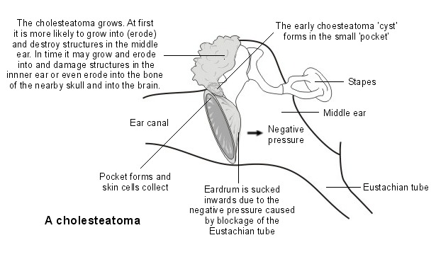
The most common cause is poor ventilation of the middle ear space, which is called Eustachian Tube Dysfunction (ETD).
Cholesteatoma can develop when the skin of the ear canal passes through a hole in the eardrum and into the middle ear space. There are several theories as to how cholesteatoma is formed or occurs in individuals.
Many theories indicate that improper functioning of the Eustachian tube often contributes to the formation of cholesteatoma.
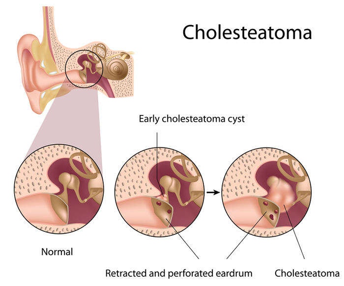
If the Eustachian tube does not open enough to equalize the pressure in the middle ear, then the negative pressure will develop behind the eardrum.
This causes the eardrum to become retracted, thus, forming a pocket. As the pocket deepens, it becomes trapped in your ear as a skin cyst or sac.
Perforated eardrum (due to infection or injuries) provide an opening for the skin of the eardrum’s outer surface to grow through. This may lead to Cholesteatoma.
Like skin tissue anywhere in the body, dead skin cells shed. As more dead skin cells shed, the sac gradually expands and cholesteatoma develops.
In other cases, skin grows around the margin of a perforation onto the middle ear.
What Are The Symptoms of Cholesteatoma?
If you previously had problems with fluid build-up and infections in the middle ear, then you may also be more likely to develop cholesteatoma.
However, the process is gradual and takes quite a long time to get developed. But, it can also be determined through its commonly occurring symptoms.
The symptoms typically start mildly but they become more severe as the cysts grow larger and begin to cause problems within one year.
A cholesteatoma usually affects only one ear. During its early stages, the common symptoms are minimal and often ignored. A person suffering from cholesteatoma may show the symptoms given below:
- Hearing loss in one ear
- The people may have ear discharge and conductive hearing loss in one or both ears.
- Ear drainage with an unpleasant smell
- Recurrent ear infections
- The sensation of ear fullness or pressure
- Dizziness
- Facial muscle weakness on the side of the infected ear
- Earache
- Ear bleeding
- Balance disruption
- Tinnitus
- History of past ear infection or fluid build-up problems
- Vertigo
- Facial muscle weakness
- Permanent hearing loss
Most of the time, the person may not know that he or she is affected by cholesteatoma until they start showing the symptoms as mentioned above.
Complications of Cholesteatoma
The accumulation of dead cells in the ear will make the bacteria and fungus to grow and survive. As a result, the cyst can become infected, causing inflammation and continual ear drainage.
Cholesteatoma is often not painful, but infection may occasionally occur, causing further pain and swelling behind the ear. In very rare cases, an infection can spread into the inner ear and brain, leading to brain abscess or meningitis.
Thus it may lead to the following complications :
- Chronic infection of the ear
- Swelling of the inner ear
- Paralysis of the facial muscles
- Meningitis, which is a life-threatening brain infection
- Brain abscesses or collections of pus in the brain
How Cholesteatoma Is Related To Hearing Loss?
- cholesteatoma and hearing loss
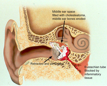
The expanding cholesteatoma sac generally causes the destruction of the eardrum and ossicles. This further causes problems in hearing.
There can be discharged from the ear, which is usually intermittent but can also be persistent. Sometimes, there is a discharge of blood.
Due to the accumulation of these skin cells, the growth can increase in size and destroy the delicate bones of the middle ear. This may affect hearing, balance, and the function of facial muscles.
Although it is a rare problem, if left untreated, it can damage the delicate structures inside the ear that are essential for balance and hearing.
It may also destroy the surrounding bone and damage the eardrum, the bones near the brain, the nerves of the face, and the bones inside the ear. Permanent hearing loss can occur if the bones inside the ear are broken.
A cholesteatoma is able to destroy the three small bones of the middle ear (the malleus, incus, and stapes, collectively called ossicles) if left untreated and the outcome of this can be a nerve deterioration, deafness, imbalance, and vertigo.
It can also affect the thin bone structure that isolates the top of the ear from the brain, as well as lay the covering of the brain open to infection with serious complications (rarely even death due to brain abscess and septicemia).
If you experience any or all of these symptoms or complications, it is advised that you immediately seek a doctor or an ENT (ear, nose, and throat) specialist for effective diagnosis and treatment.
How Is Cholesteatoma Diagnosed?
Cholesteatoma is generally detected by examining the ear and finding the disease. However, the doctor may suspect the disease when some or all of the following are present:
- The gradual loss of hearing
- Discharge of especially blood from the ear
- History of past ear infection or fluid problems
Your audiologist may refer to the following diagnostic methods:
1. Otoscopic exam
The doctor will check your ears with the help of an otoscope to determine whether you have a cholesteatoma. This medical device allows the doctor to see if there is a sign of a growing cyst.
Specifically, they will look for a visible deposit of skin cells or a large mass of blood vessels in the ear.
2. CT Scan
Mostly treatments include imaging such as CT Scan that helps to define where cholesteatoma is located. This is required when there is no obvious sign of cholesteatoma.
CT scans will be used if you are showing symptoms such as dizziness and facial muscle weakness. It is a painless imaging test that is used to captures images from a cross-section of your body.
The scan allows your doctor to see inside your ear and skull. This can help them better visualize the cyst or rule out other possible causes of your symptoms.
3. Ear Microscopy
The typical findings here are a defect of the tympanic membrane, white epithelium scales, and eventually bone erosion (progressive destruction) of the ear canal wall, close to the eardrum.
4. Hearing Test (Audiogram)
Typically, cholesteatomata patients suffer from conductive hearing loss, i.e., a hearing disorder that only affects the outer ear.
If the cholesteatoma is so far advanced that the inner ear is already affected, a so-called sensorineural hearing loss is present.
Thus the hearing test is essential prior to surgical treatment in order to know about the extent of hearing loss a person is suffering.
Other than this, an MRI might also be required, if needed. Since there are chances of cholesteatoma to reoccur in a person, it is important to keep a follow up regularly with your doctor.
Treatment of Cholesteatoma
Cholesteatoma Surgery is the only option as the cyst needs to be removed surgically in order to prevent complications. It doesn’t go away naturally.
They usually continue to grow and cause additional problems. Although it can be managed in different ways, the removal of the dead skin cells or cyst may require surgery.
Prior to surgery, the ENT specialist may need to carefully clean your ear and prescribe medications to help stop the drainage.
As after cleaning the drainage and infections, your audiologists will be able to better analyze the growth traits of the cyst and make a plan for surgical removal.
Thus, the treatment of Cholesteatoma is completed in two steps:
- Cleaning of the infections or fluids from the ears
- Cholesteatoma surgery
1. Cleaning the Cysts and Infections
A regimen of antibiotics, ear drops, and careful cleaning of the ear will most likely be prescribed to treat the infected cyst, reduce inflammation, and drain the ear.
The medication may be taken by mouth or applied directly to the ear, or sometimes both may be required. Thus, it is advised that you keep the ear dry while treating the infections.
The specific type of surgery depends exactly on what part of the ear is involved with cholesteatoma.
Until the doctor cleans your ears and inspected the entire tympanic membrane, Cholesteatoma cannot be diagnosed.
2. Cholesteatoma Surgery
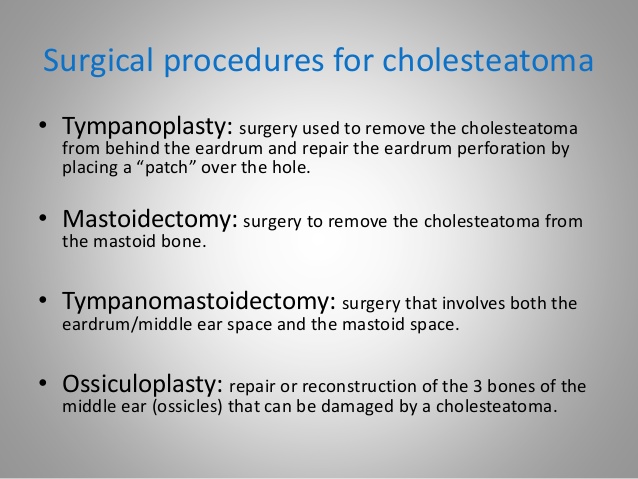
In most cases, outpatient surgery is required in case of treating Cholesteatoma. The surgery is done under general anesthesia.
After the initial surgery to remove the cyst, a follow-up surgery is required to reconstruct any damaged portions of the inner ear and make sure that the cyst has been completely removed is often necessary.
This typically involves some form of mastoidectomy which may or may not involve removing the posterior ear canal wall and the ossicles.
During surgery, the mastoid is opened and the eardrum is elevated and upon removing all diseases, the eardrum is then rebuilt with a graft obtained from the lining of muscle behind the ear.
If there is a defect in the ear canal adjacent to the eardrum, then it may be rebuilt with cartilage. One of the most effective strategies for achieving the primary aim of cholesteatoma surgery (complete removal of cholesteatoma) is the removal of the canal wall.
Post-Surgical Procedures of Cholesteatoma
After the removal of Cholesteatoma, you must attend the follow-up appointments to estimate results and ensure the cyst hasn’t come back. If the cyst broke any bones in your ear, you’ll need a second surgery to repair them.
Follow up is necessary because even after careful microscopic surgical removal, cholesteatomas may recur. Such recurrence may arise many years, or even decades, after treatment.
After surgery, some people experience temporary dizziness or taste abnormalities. These side effects almost always resolve themselves within a few days.
It is not possible to protect yourself from Congenital cholesteatomas, but you should aware of the changes you may experience due to cholesteatoma.
You can prevent cholesteatomas later in life by treating ear infections quickly and thoroughly. However, cysts may still occur.
It’s important to treat cholesteatomas as early as possible to prevent complications. Call your doctor as soon as possible if you believe you have a cholesteatoma.
Conclusion
If a cholesteatoma has become large and complex before it is identified then it might lead you to permanent hearing loss and it can also result in vertigo or imbalance.
But these complications are often rare if the cholesteatoma cyst is caught and remove early.
You can purchase the latest hearing aids at a fair price through HearingSol, If you have a query regarding Cholesteatoma and Hearing Loss, feel free to call us at +91-9327901950. We are always here to help you.