 Reviewed by Mr. Ranjeet Kumar
Sr. Audiologist, Speech Therapist & Cochlear Implant Specialist, BASLP on
Reviewed by Mr. Ranjeet Kumar
Sr. Audiologist, Speech Therapist & Cochlear Implant Specialist, BASLP on
There are over 600 muscles in the human body, which together account for about 40 percent of the body weight. Humans have well-developed muscles in the face that permit a large variety of facial expressions.
Through these muscles, we use to show expressions like happiness, surprise, disgust, anger, fear, and other emotions, they are thus, an important means of non-verbal communication.
As these muscles are helpful in playing such an important part like giving expressions, other muscles in our body have the same importance for different activities.
If you need any assistance or have a question about Head And Neck Muscles Functioning & Location, you can consult our HearingSol experts with your problem, feel free to call us on +91-9327901950. We are always here to help you.
In this article, we are going to discuss Head and Neck muscles, how many they are, how they work, how important they are, and all the related things.
Neck Muscles Functioning
The Neck is an area between the head and the rest of the body and it is built of different tissues and organs, including many skeletal muscles.
The main function of the neck is to allow movements of the neck or head, providing structural support to the head.
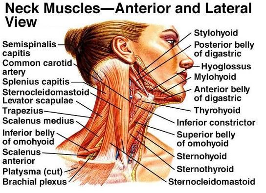
- Neck Muscles
Muscles of the neck are divided into groups according to their working:
1. Superficial Neck Muscles
It is the most external muscle which includes:
- Platysma – It is a wide flat, superficial neck muscle spreading from the lower part of the face to the upper thorax. It is a paired thin muscle arising from the upper parts of the shoulders and inserting into the mouth. It involves in depress and wrinkles skin of the lower face and the mouth.
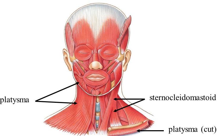
- Sternocleidomastoid – It is a paired muscle that is located on each side of the neck. It origins from the manubrium of the sternum and inserts on the superior nuchal line of the occipital bone.
2. Scalene Muscles
The scalene muscle is a triple paired muscle in the sideward part of the neck. It is divided into three parts:
-
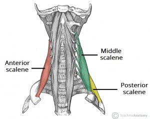
- Scalene Muscles
- Anterior Scalene – It is a deep muscle of the sideward part of the neck and lies behind the sternocleidomastoid. It originates from the anterior tubercles and inserts into scalene tubercles on the inner border of the first rib. It helps in flexing or bending the neck.
- Middle Scalene – It is the largest and longest of Scalene muscles. It is located between the anterior and posterior scalene muscle in the neck. It originates from the anterior tubercles of the transverse processes of the 2nd to 7th cervical vertebrae and inserts onto the upper surface of the first rib.
- Posterior Scalene – It is the smallest scalene muscle situated most deeply. It originates from the posterior tubercles of the transverse processes 5th to 7th cervical vertebrae and inserts on the outer surface of the second rib.
3. Suprahyoid Muscles
The suprahyoid muscles are a group of neck muscles that are located above the hyoid. It consists of three types of muscles
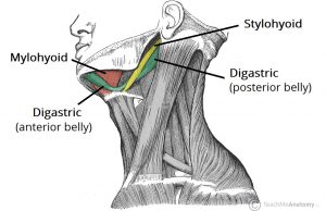
- Suprahyoid Muscles
- Digastric – It is located below the mandible and consists of two muscular bellies that are Anterior and posterior bellies. These two are connected by an intermediate tendon. The digastric muscle opens the jaw when the masseter and the temporalis are relaxed.
- Mylohyoid – It is a paired muscle of the neck that extends from the mandible to the hyoid bone. It also helps in forming an oral diaphragm. It originates from the mylohyoid line on the internal surface of the mandible and inserts onto the body of the hyoid bone.
- Stylohyoid – It is a neck muscle stretching between the base of the skull and the hyoid bone. It originates from the styloid process of the temporal bones and inserts on the lesser horn of the hyoid bone. It helps in pulling of hyoid bone backward and upward.
4. Infrahyoid Muscles
The infrahyoid muscle is located below the hyoid bone and involves in repressing it. It includes:
-
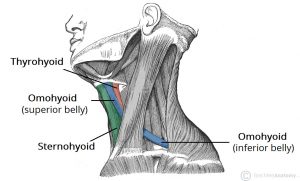
- Infrahyoid Muscles
- Omohyoid – It belongs to the Infrahyoid muscle and depresses the hyoid bone. It originates from the upper border of the scapula and inserts into the inferior part of the hyoid bone.
- Sternohyoid – It is a thin neck muscle extending between the sternum and the hyoid bone. It originates from the manubrium of the sternum and inserts into the body of the hyoid bone. Its main work is depressing the hyoid bone.
- Sternothyroid – Sternothyroid muscle extends between the sternum and the thyroid cartilage. It originates from the manubrium of the sternum and inserts into the thyroid cartilage of the larynx. Its work is to depress the thyroid cartilage.
- Thyrohyoid – It is located in the neck region between the thyroid cartilage and the hyoid bone. It originates from the oblique line of the thyroid cartilage and inserts into the greater horn of hyoid bone. Its main function is to pull the hyoid bone downward and bring the hyoid bone and the thyroid cartilage closer together.
5. Prevertebral Muscles
This muscle is situated anterior to the vertebral column. It includes:
- Prevertebral Muscles
- Longus Capitis – It is a prevertebral muscle of the neck that is located anterior to the vertebral column. It originates from the anterior tubercles of the transverse processes of the 3rd to 6th cervical vertebrae and inserts into the inferior surface of the occipital bone. Its main function is flexion and rotation of the head.
- Longus Colli – It is an extending muscle between the atlas and the thoracic vertebrae. It is responsible for the flexion of the head and neck. It originates from the transverse process of the 2nd to 5th cervical vertebrae and inserts into the anterior tubercles of the atlas.
- Rectus Capitis Anterior – It is a short muscle located in a vertebral column, stretching between the atlas and the base of the skull. It originates from the base of the transverse process of the atlas and inserts into the surface of the occipital bone. It is responsible for the flexion of the neck at the atlanto-occipital joint.
- Rectus Capitis Lateralis – It originates from the upper surface of the transverse process of the atlas (C1) and inserts into the surface of the jugular process of the occipital bone. It helps in lateral flexion of the neck and also in the stabilization of the atlantooccipital joint.
6. Suboccipital Muscle
-
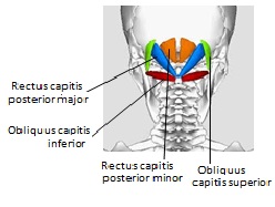
- Source: niagarachiroandmassage.ca
These muscles are located in the upper posterior part of the neck and below the occipital bone. These are also the deep muscles of the back. They include:
- Obliquus Capitis Inferior – It is a suboccipital muscle that acts as the atlantoaxial joint. It originates from the spinous process of the axis and inserts into the transverse process of the atlas. It is involved in the rotation of the head and neck.
- Obliquus Capitis Superior – It is a muscle that acts as the atlantooccipital joint. It originates from the transverse process of the atlas and inserts into the half of the inferior nuchal line on the surface of the occipital bone. It helps in extending the head and also helps in flexing the head to the ipsilateral side.
- Rectus Capitis Posterior Major – It originates from the spinous process of the axis and inserts into the inferior nuchal line on the surface of the occipital bone. It is involved in the ipsilateral rotation of the head and extension of the head.
- Rectus Capitis Posterior Minor – It also acts as the atlantooccipital joint. It originates from the tubercles on the posterior arch of the atlas and inserts into the external surface of the occipital bone. Its main function is the extension of the head.
That is all the muscles that include in Neck muscle functioning. Now its time to discuss Head Muscles and their functions.
Muscles of the Head
The head muscles can be grouped into two categories, Masticatory muscles which are derivatives of the first pharyngeal arch, and Facial muscles which are derivatives of the second pharyngeal arch.
These two groups of head muscles are supplied by two different cranial nerves because of their different Embryological development. Most muscles of the head belong to the facial muscles and are innervated by the facial nerve. While the masticatory muscles are innervated by the Mandibular nerve.
Let’s discuss other components of these two categories of head muscles
1. Masticatory Muscle
This muscle is responsible for the chewing movements of the lower jaw. It originates on the skull and inserts into the mandible, thereby acting upon the movements of the lower jaw at the Temporomandibular joint.
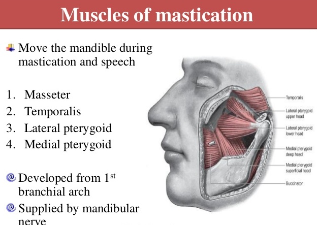
- Masticatory Muscle
There are 4 masticatory muscles on both sides of the head. These are:
- Masseter – The masseter is a masticatory muscle that lifts up the movement of the lower jaw. This muscle originates from the Zygomatic arch and inserts into the masseteric tuberosity of the mandible. It is thick and quadrangular in shape.
- Temporalis – The temporalis is involved in the elevation and retraction of the lower jaw. It is a wide, fan-shaped muscle on each side of the head that covers most of the temporal bone. It is covered by temporal fascia.
- Lateral pterygoid – The lateral pterygoid muscle pushes the mandible forward. And, while by contraction on one side, this muscle pushes the mandible to the opposite side. It is a thick, short muscle located in the infratemporal fossa.
- Medial pterygoid – The medial pterygoid helps in the elevation of the mandible acting as a synergist to the temporalis and masseter muscles. It is involved in many movements of the mandible. It is located in the infratemporal fossa.
The masticatory muscles are innervated by the Mandibular nerve. Now let’s discuss facial muscle:
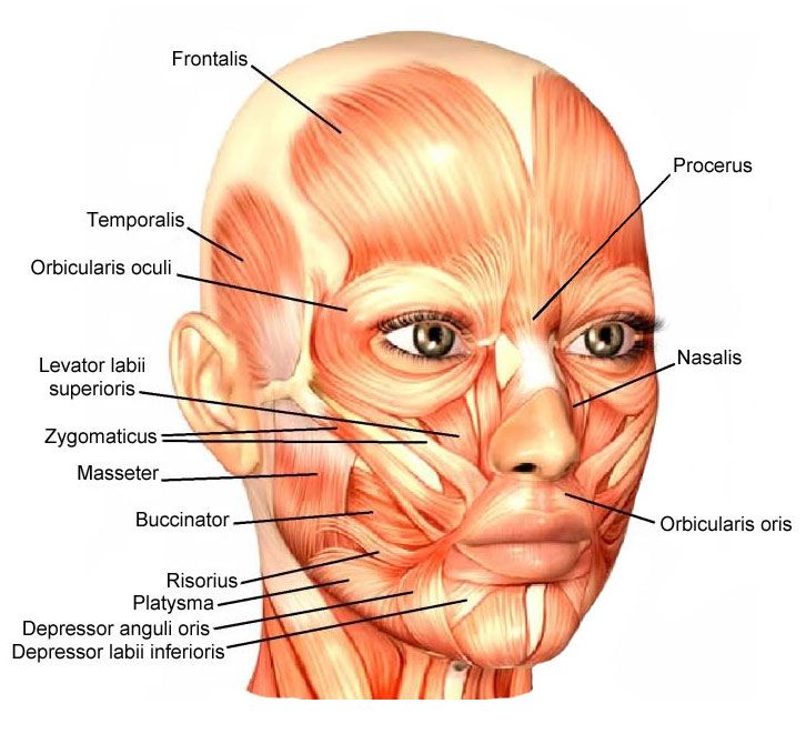
2. Facial Muscle
These muscles are situated under the subcutaneous tissue. Facial muscles are responsible for the movements of skin folds, different facial expressions. It originates from the bones of the facial skeleton and inserts it into the skin.
These muscles are normally grouped around the natural openings of the face like eyes, nose, or mouth and are responsible for the closing or widening of these openings.
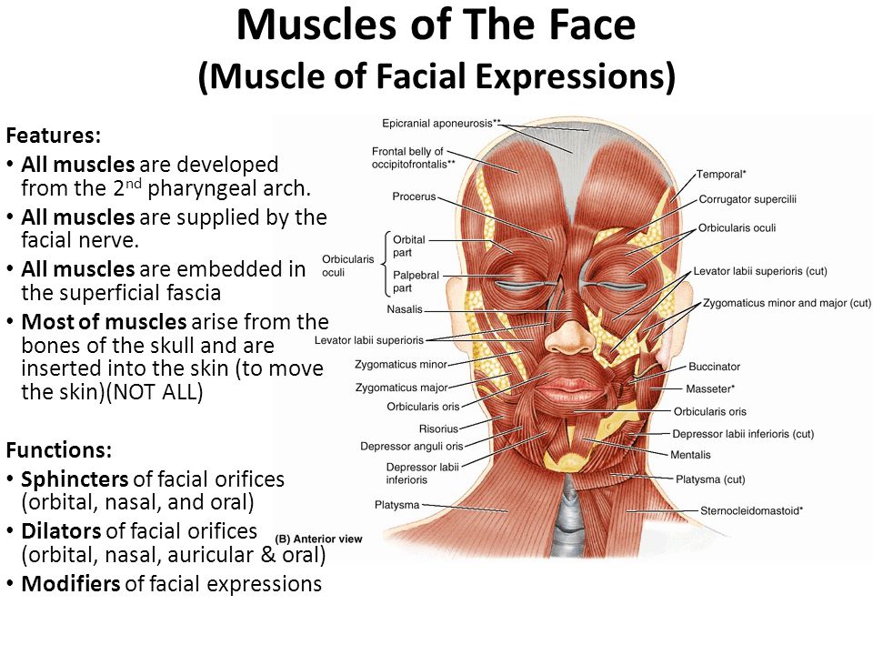
- Facial Muscles
Facial muscles can be divided into 3 categories. These are –
(A). Muscles of the Orbital opening (Eyes),
(B). Oral opening (Mouth)
(C). Nose opening.
Let’s discuss all 3 step by step:
(A) Facial Muscles Around The Orbital Opening (EYES) Include
- Corrugator Supercilii – It is a small, narrow facial muscle located at the end of the eyebrow. It originates from the superciliary arch and inserts into the skin over the middle of the eyebrow. It pulls the skin of the eyebrow downward and produces wrinkles of the forehead.
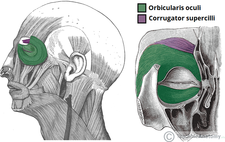
- Orbicularis Oculi – It is a circularly shaped muscle around the opening of the eye. It is involved in the closing of the eyelid. It consists of 3 parts – The orbital, the palpebral, and the lacrimal. The Orbital part lies around the orbit opening, the palpebral part is situated in the upper and lower eyelids, and the lacrimal lies the most profoundly.
(B) Facial Muscles Around The Oral Opening (MOUTH) Include
- Buccinator – It is involved in forming the anterior part of the cheek and the sided wall of the oral vestibule. It origins From the alveolar process of the maxilla and at the region of 1st and 2nd molar teeth and inserts into the angle of mouth passing out into the fibers of the orbicularis oris muscle.
- Depressor Anguli Oris – It is responsible for pulling the angle of the mouth downward and enabling facial expression associated with sadness. It originates from the base of mandible and inserts into the skin of the angle of the mouth.
- Depressor Labii Inferiors – It is a muscle of the chin area that pulls the lower lip downward and forward. It originates from the oblique line of the base of mandible and inserts into the skin of lower lips. It participates in producing facial expressions of diligence.
- Levator Anguli Oris – It is used in lifting the angle of the mouth. It origins from the canine fossa on the anterior surface of the maxilla and inserts into the skin of the angle of the mouth. It participates in creating a smile.
- Levator Labii Superiors – It is used to lift the upper lip. It origins from the infra-orbital margin of the maxilla and inserts into the skin of the upper lip. It helps in producing facial expressions of worry, anxiety.
- Facial Muscles Around The Oral Opening
- Mentalis – It origins from the incisive fossa on the alveolar process of the mandible and inserts into the skin of the chin. It is located in the chin area and produces small dimples in the chin.
- Orbicularis Oris – It is located in the thickness of the lip around the oral opening. It is used in the closing of the mouth. It has two main parts that consist of longitudinal fibers and circular fibers. The longitudinal fibers origins below the skin around the mouth opening and inserts inside the tissue of the lips. While the origins of the circular fiber from the angles of the mouth and insert within the skin of the corner of the mouth.
- Risorius – It is located sidelong to the mouth opening which pulls the angle of the mouth laterally. It originates from the masseteric fascia and inserts into the skin of the angle of the mouth. It helps in producing facial expressions of pleasure and laughter.
- Zygomaticus Minor – It is located medially to the zygomaticus major and belongs to elevators of the lip. It originates from the surface of the zygomaticus bone and inserts into the skin of the upper lips. It helps in pulling the upper lip backward, upward, and outward.
- Zygomaticus Major – It is involved in creating an expression in the human face known as a smile. It originates from the zygomatic arch and inserts into the angle of the mouth. It also helps in the formation of cheek dimples in some individuals.
(C) Facial Muscles Around The Nose Opening Include
- Compressor Nerium Minor – It is a small muscle located in the nose region and it is present in nearly more than half of individuals, seems to play role in decreasing the nasal opening, narrowing the nostrils. It originates from the anterior part of the lower cartilage of the nose and inserts into the skin near the margins of the nostrils.
- Dilator Naris Anterior – It encircles the nostril and helps in widening it. It origins from the upper cartilage of the nasalis and inserts into the skin at the caudal margin of the lateral crus.
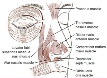
- Levator Labii Superiors Alaeque Nasi – It helps in widens the nostrils and elevates the upper lips and the wing of the nose that provides the facial expression of snarling. It originates from the frontal process of maxilla and inserts into the skin of the nostrils and upper lip.
- Nasalis – It is a paired facial muscle that is located in the area of the nose. It consists of two parts – The Transverse part which compresses the nostrils and The alar part which widens the nostrils.
- Procerus – It is an unstable facial muscle that is located between the forehead and the dorsum of the nose. It originates from the midline of the nasal and inserts into the skin of the lower part of the forehead between the eyebrows. It involves producing a transverse fold across the root of the nose.
Outlook
However, every part of our body is important for us because every part plays a different and important role. But Head and neck muscles are one of the most important parts of our body.
We mentioned it “most important” because they are sensitive and can’t be replaced if any untreatable harm happened to them. And definitely, you can’t live without them either.
For instance, people have two kidneys but most people survive with just one. The appendix is also an example of a body part that can be totally removed and we can live without it. In several cases of appendicitis, when the appendix becomes inflamed, it has to be surgically removed.
But in case of head or neck muscles, if any harm or disease happens to any one of the two, there is only one method and that is Treatment only. You cannot remove these parts nor you can replace them.
That is why it is suggested that if you feel any kind of discomfort or any other type of issue in head and neck muscles, do not ignore it and treat it as soon as possible because if this problem increased it can affect your working, relationships and many other daily life activities.
If you need any assistance or have a question about Head And Neck Muscles Functioning & Location, you can consult our HearingSol experts with your problem, feel free to call us on +91-9327901950. We are always here to help you.
Read More: Know the location and Functioning of Head & Neck Bones